02.23.23
How to Prepare for your Duplex Ultrasound in Rogers
Step one in treating your health concerns is identifying the issue itself. As the saying goes: you can’t clear your veins until you map them with sonic imaging technology. That’s a phrase, right?
Well, people have a lot of questions about vascular testing—be it an ultrasound, an x-ray, or an ankle-brachial index test (ABI). The single most common question is, “how do I prepare?” So, what do you need to do?
The Ozark Regional Vein & Artery Center team thought a quick guide to prepping for a duplex ultrasound in Rogers could be a help to patients. Whether you’re neighbors coming to see us from Fayetteville and Bentonville or long-distance travelers from Springfield, MO, and Houston, Texas, we’re here to help!
So, in this blog, we’re going to cover how to prepare for vascular screening, what to look for in a vascular imaging provider, and then discuss the technology itself and what you should know. That way, you can go into your ABI or duplex ultrasound in Rogers comfortably and with confidence.
How should I prepare for vascular testing?
Fast before your screening
To prepare for a vein screening, don’t eat for 6-8 hours before your vein screening. This is to help us get the clearest sense of the actual state of your veins and arteries. Eating a big meal before your visit will throw off our ability to gauge the health of your blood vessels.
Besides restricting your eating and drinking, there is no other special preparation for a venous ultrasound!
Dress comfortably
That being said, we recommend wearing comfortable, loose-fitting clothing and leaving your jewelry at home. Since you may need to remove clothing or jewelry in the screened area, this policy helps to save you time. And who wouldn’t want to be as comfortable as possible during an ultrasound?
Take a deep breath
Lots of patients come to their vascular test in Rogers anxious and afraid of their results. That’s entirely understandable. Vascular healthcare can be very frightening, especially if you’re unsure what you’re dealing with. But make sure to take a deep breath. You’re not in this alone, and there’s no vascular condition that a little hard work can’t improve.
After all, that’s what the Ozark team is here to do! So remember: identifying the problem isn’t the end of your life; it’s the first step on your road to recovery.
Why are we talking about Duplex Ultrasound?
As a quick overview, the Ozark Regional Vein & Artery Center offers a variety of screening technology to test your veins & arteries for vascular disease or buildup. One of our most effective tools is the duplex ultrasound. This test uses sound waves to produce images of the veins & arteries in the body.
That sonic map is how providers at the Ozark Regional Vein & Artery Center use screening to search for blood clots in leg veins and elsewhere. It’s the perfect tool for identifying deep vein thrombosis (DVT), venous insufficiency, and arterial health concerns. While it’s more commonly used for arterial health screening, we use it to get a comprehensive view of your vascular health.
While it’s not the only test we offer, it’s commonly performed and is a great basic treatment to give a rundown of. Most vascular imaging will have the exact same preparation requirements as a duplex ultrasound in Rogers.
What should I look for in vascular screening providers?
At Ozark Regional Vein & Artery Center, our priority is providing last vascular healthcare solutions. That means not only providing world-class vascular treatment, but also the screening and imaging services necessary to give you peace of mind.
Unlike hospital appointments, our team can get patients in for testing and provide them with results within 48 hours. We offer a complete suite of testing services and run a full vascular lab with duplex ultrasound in Rogers that can quickly get the answers you need.
Advanced Technology
Our artery center offers angiograms, duplex ultrasound, carotid duplex, and ankle-brachial index or ABI tests. These are the most common and valuable arterial vascular tests in the medical field.
We have several noteworthy devices for imaging, including the OEC Elite C-Arm and Phillips IVUS Ultrasound machine, to name a few.
The OEC Elite C-Arm is a world-renowned surgical X-Ray device that optimizes procedures Dr. Stout performs. The Phillips IVUS ultrasound machine is another gold-standard technology our team uses to analyze the arterial system and aid in placing stents, angioplasties, and more.
Expert Providers
Dr. Stout and Dr. Haney are the two leading medical providers here at the Ozark Regional Vein & Artery Center. Dr. Stout is the resident vascular surgeon at our facility, and Dr. Haney is our founder and medical director. They have over 50 years of combined experience providing quality vein & artery care to patients across the country.
So, not only is there no one more qualified to perform your blood circulation test in Rogers, there are no medical providers better equipped to care for your vascular health.
What is vascular imaging and how does it work?
We’ve gone over how to prepare for your vascular imaging test and what to look for in a vein screening provider, but there’s a lot more to know about the technology itself. After all, lots of people haven’t gotten to learn about x-rays and ultrasound devices.
Reasonably, this leaves new patients with a lot of misconceptions and questions about imaging technology. If something’s going to peer around inside our bodies, we want to know how it works!
Ultrasound Test
At its most basic level: sonography (ultrasound imaging) is a noninvasive test that helps diagnose and treat medical conditions. It produces pictures of the inside of the body using sound waves. It uses a small probe called a transducer and gel placed directly on the skin. High-frequency sound waves travel through the gel into the body.
A computer uses those sound waves to create an image. Because ultrasound captures images in real-time, it can show the structure and movement of the body’s internal organs and blood flow.
Ankle-Brachial Index Test
The ankle-brachial index (ABI) is a non-invasive screening tool that measures variances in blood pressure. Specifically, it compares the blood pressure in your lower extremities as your heart is beating (systolic blood pressure) with that of your upper body & extremities.
Ankle-brachial index gets its name from the fact it’s easiest to test for that pressure along the ankle. Wide variation between the two numbers is an immediate indication there is some degree of obstruction or weakness in the veins and arteries in your legs.
Angiogram
An angiogram is an X-ray procedure that diagnostically identifies blockages in the arterial system. An angiogram does this using X-rays taken after the injection of a contrast agent (iodine dye) into the bloodstream.
Angiograms are typically performed while you are sedated. The procedure usually lasts 20-25 minutes but can take longer. It will ultimately depend on the complexity of the issue and the resolution of the first round of x-rays.
What are some common uses of duplex ultrasound in Rogers?
The most common reason for a venous ultrasound exam is to search for blood clots, especially in the veins of the leg. This condition is known as deep vein thrombosis (DVT). These clots may break off and pass into the lungs, where they can cause a dangerous condition called pulmonary embolism. If we find a blood clot early enough, proactive treatment can prevent it from worsening.
At the Ozark Regional Vein & Artery Center, we commonly use vascular imaging to determine the cause of varicose veins. It allows us to identify the exact location of any varicose veins and pinpoint the precise location of the cause.
Likewise, a duplex ultrasound in Rogers is an ideal tool for identifying vascular conditions of the deep veins or arteries. It is able to create a much more extensive map of your blood vessels, allowing for far more thorough diagnostic support than other testing.
Whether a weak valve or some form of a clot, a vascular screening helps us get the whole picture. That’s a little radiology pun for you.
Some other common uses of this technology include:
Surgical Support
Aid in guiding a needle or catheter placement into a vein. Sonography can help locate the exact site of the vein and avoid complications, such as bleeding or damage to a nearby nerve or artery.
Monitoring Recovery
Clots can form suddenly, even after successful treatment. There are a variety of reasons, many of which are out of your control. If your vascular provider places a line or stent in a leg or arm vein, there is a potentially higher chance of developing a clot around it.
In all these instances, regular screening is vital to making sure you’re healthy, and your vascular system isn’t at risk.
Duplex ultrasound or another blood circulation test will help your provider see and evaluate:
- blockages to blood flow (such as clots)
- narrowing of vessels
- tumors and congenital vascular malformations
- reduced or absent blood flow to various organs, such as the testes or ovary
- increased blood flow, which may be a sign of infection
How does duplex ultrasound in Rogers work?
Ultrasound imaging uses the same principles as the sonar that bats, radar, and dolphins use. When a sound wave strikes an object, it bounces off of it. By measuring these echo waves, it is possible to determine how far away the object is and its size, shape, and consistency. This includes whether the object is solid or filled with fluid.
Medical professionals can take advantage of ultrasound technology to detect changes in the appearance of organs, tissues, and blood vessels. This is how ultrasounds can be used to detect blood clots, abnormal growths, or even tumors.
During an ultrasound exam, the transducer sends out sound waves and records the echoing (returning) waves. When your provider presses the transducer against your skin, it sends small pulses of soundless, high-frequency sound waves into the body.
As the sound waves bounce off internal organs, fluids, and tissues, the sensitive receiver in the transducer records tiny changes in the sound’s pitch and direction. A computer instantly measures these sound’s waves and displays them as real-time pictures on a monitor.
It’s practically magic. But it’s not magic. It’s science.
How is the vascular imaging procedure performed?
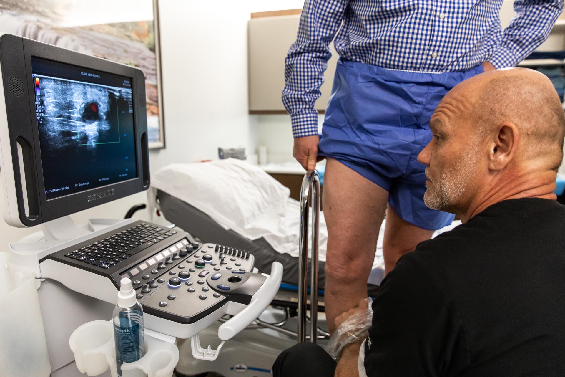
While many medical facilities that perform vein tests ask you to lie down during screening, the Ozark Regional Vein & Artery Center always tries to keep you standing to perform a screening on your legs and lower body. Laying down means your body isn’t having to work as hard as it normally would because blood isn’t having to pump against gravity. That gives us a less accurate picture of what your body’s blood vessels are actually dealing with day to day.
Think of it this way: if you’re a restaurant trying to determine how much food you can serve during a lunch rush, you wouldn’t want to do it on a day when half the servers are on vacation. You want to get a picture of your body working like it normally does.
The Process
Your Ozark Regional provider will apply a water-based gel to the area being screened. This helps the ultrasound transducer make secure contact with the body. It also helps eliminate air pockets between the transducer and the skin that can block the sound waves from passing into your body.
Your provider will sweep the ultrasound transducer over the areas of concern, repeating the process multiple times to get a more complete picture. They may also angle the sound beam from a different location to better see an area of concern.
This ultrasound examination is usually completed within 20-30 minutes. More complex exams may take more time.
What will I experience during and after the procedure?
Most vein screening exams are painless, fast, and comfortable. There is usually no discomfort from pressure as they press the transducer against the area under examination.
If scanning is performed over an area of tenderness, you may feel pressure or minor pain from the transducer.
Once the imaging is complete, your provider will wipe off the clear imaging gel from your skin. If it’s a point of concern, the gel does not usually stain or discolor clothing, but tossing an article of clothing into the wash should prevent damage to your clothing.
After an ultrasound exam, you should be able to resume your normal activities immediately.
Who interprets the results, and how do I get them?
Dr. Haney, Dr. Stout, and our entire vein care team are versed in performing vascular screenings and can provide you with comprehensive explanations of your results and what to expect post-screening.
Dr. Haney & Dr. Stout will always review your results, meaning that they will be the ones to walk you through the more complex implications of your vascular condition. This is when we determine whether you’re dealing with a vein concern, artery concern, or both.
Related: Veins, Arteries, and Which Doctor is Right for You
It’s worth noting that you may require a follow-up screening. This is normal and nothing to worry about. Sometimes a follow-up exam further evaluates a potential issue with more views or a special imaging technique. It may also see if there has been any change in an issue over time.
Likewise, follow-up exams are often the best way to see if treatment is working or a problem needs attention.
Turn to the Ozark Regional Vein & Artery Center for your vascular screening needs.
For vascular imaging and/or a duplex ultrasound in Rogers, come to the Ozark Regional Vein & Artery Center. Our experience and growing suite of care options allow us to guide you toward lasting wellness solutions for a happier, healthier life.
We are a premier practice in Northwest Arkansas for all the highest-quality vascular treatments available. Dr. Haney, Dr. Stout, and the expert staff have over 75 years of combined experience in the industry. Patients come to us from across the country—from Fayetteville & Bentonville to Houston, Texas, & Springfield, Missouri—to ensure they receive the best concierge-level care.
After all, helping people is what we do. And it is our mission to provide the people of Arkansas and beyond with the absolute best care possible.
Take the first step on the road to recovery with our Virtual Vascular Screening Tool, or schedule a consultation. We also hold regular free screening events.
Sign up for our newsletter
Want to be in the know about your vascular care? Join our newsletter ⬇️ It’s a great way to stay notified of our free public events and other practice specials!